The Charity supports the cardiology service at King’s by funding cutting-edge equipment and the team carrying out vital research. King’s has a large, active team investigating many aspects of heart disease.
Finding innovative treatments
Your support has helped to fund three research fellows running a programme seeking new ways of treating heart valve disease. They are using the latest imaging techniques and 3D printing to create models of heart valves, which are then put into a special machine – also funded by donations – that simulates blood flow. Consultants can then use this method to practise surgical techniques and test which devices work best for different types of disease.
As the average age of the UK population increases, so does the prevalence of heart disease. This research, and the new ultrasound scanner at King’s, means consultants can offer more minimal access treatment rather than open-heart surgery.

Supporting safer surgery
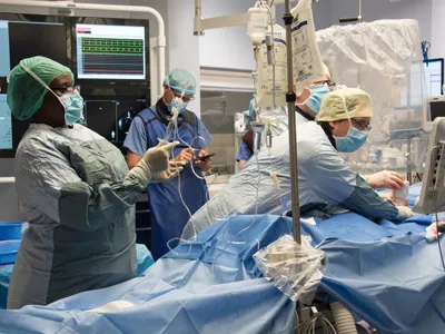
Our support has provided a new ultrasound scanner, which makes heart surgery safer and quicker because surgeons can reach less accessible arteries more easily.
When surgeons put devices into the heart, such as replacement valves, it’s usually done through an artery in the groin because it’s one of the biggest arteries in the body. But it’s buried deep within the upper thigh and can be hard to locate. Thanks to the new scanner, the artery can be visualised on screen.